A Patient’s Guide to Heel Pain …
What You Need To Know
What are the symptoms of heel pain?
The typical symptoms of heel pain are pain on the inside of the heel when you stand or walk after periods of rest or inactivity; especially pain with the first step in the morning. The pain lessens after walking for a while. Typically in the afternoon the heel hurts whether you rest or walk on it. The pain can vary from mild to debilitating and can last from a few weeks to many years. The pain can be centered just under the heel or it can extend across the arch of the foot. The pain can appear to be cured, at times, only to return months or years later. You may have noted that shoes with a bit of an elevated heel or walking with your feet turned inward helps to relieve the pain. Plantar fasciitis is uncommon, but not unheard of, in anyone younger than 30.
What is going on here?
 To understand heel pain you first need a quick lesson in anatomy. There is a thick band or belt of tissue that extends from your toes to your heel and is just under the skin. The band is called the plantar fascia or PF. At the front of the foot it attaches to all five toes and at back it attaches to the heel bone, the calcaneus, with a 1 inch wide attachment. The purpose of the plantar fascia is to provide stability to the foot when you lift your heel for walking, running and climbing. It is possible to do more of these activities then your plantar fascia can handle in any given time period in pain results. So the first and simplest cause of heel pain is overuse were the plantar fascia hurts at its weakest point, the attachment site on the heel bone. We can easily see this visually in the office with the diagnostic ultrasound machine which allows us to measure the thickness of the plantar fascia at the heel attachment site. A normal plantar fascia is 3-4 mm thick and the thickness of a painful plantar fascia is typically six or more millimeters thick.
If the overuse and over-pull of the plantar fascia goes on for a long period of time, some say 18 months or more, a spur can develop on the heel bone which you can visualize with an x-ray. Most authorities believe that the heel spur is not the cause of heel pain. People can have a huge heel spur with no pain while others can have tremendous pain without a heel spur. I believe that the heel spur is a marker to indicate that the process has been going on for almost 2 years or more. These people will probably require more treatment than just a cortisone injection.
Fasciitis vs. Fasciosis
The terms fasciitis and fasciosis look suspiciously similar but they refer to very different things. The “-itis” ending of fasciitis gives a clue to its meaning: inflammation. During the early stages of this condition where the over-pull or overuse of the plantar fascia is in its beginning stages the condition is inflammatory. Anti-inflammatory pills and cortisone injections are very useful in this stage. As time passes the condition morphs into a chronic pain phase where we refer to it as fasciosis. Anti-inflammatory pills and cortisone injections are not effective during the fasciosis phase. If you have had a cortisone injection for your “plantar fasciitis” that only lasted for a few days you are probably in the “fasciosis” stage.
What causes the spur?
 The cause of the spur has been an intriguing question that does not have a clear answer. The best explanation I have heard is that the spur forms as a reaction to an accumulation of micro-fractures to the heel bone. These micro fractures, like all fractures, heal by having the tissue covering the bone, the periosteum, produce more bone which will, in effect, glue the bones together. With chronic plantar fasciitis, or fasciosis, there are tens of thousands or hundreds of thousands of little fascial tugs on the heel bone that produce tens of thousands or hundreds of thousands of microscopic heel bone fractures. The extra bone produced to heal the microscopic heel bone fractures is visualized as the spur. Although it may feel like it, the spur does not point downward towards the ground
When do you suspect that there might be a stress fracture?
Long standing over-pull and overuse of the plantar fascia can also lead to a stress fracture of the heel bone. This is not common but it can occur. I am suspicious of a stress fracture if squeezing the heel bone between the palms of my hand produces pain on the affected side but not the unaffected side. Regular x-rays taken in the office can give a clue that you might have a stress fracture. Confirmatory tests for a stress fracture often need a bone scan or an MRI both of which are done at a radiology center.
Bone scans are very useful tools. A radioactive dye is injected in the arm and an x-ray scan is taken of the heel bone three hours later. A “hot” scan which shows a lot of activity in the heel bone is diagnostic for a stress fracture. An example of a “hot” scan is attached the right heel has a very hot spot indicating that something is going wrong. It could either be cancer, an infection or a heel stress fracture, depending on the symptoms of the patient. In this case, it was a heel stress fracture.
As useful as bone scans are, they are being replaced with MRIs as the cost of the MRIs have decreased. MRIs can also get useful information about the plantar fascia and the surrounding tendons that you cannot get with a bone scan. I cannot remember the last time I ordered a bone scan to evaluate the possibility of a stress fracture in the heel. If the MRI shows you have a stress fracture expect to be in a walking cast for 3-6 weeks to allow the foot to heal.
Quick summary so far
1) Plantar fasciitis produces pain with the first step after resting
2) The most common cause of pain is inflammation of the fascia at the heel insertion
3) Long standing plantar fasciitis leads to a non-inflammatory chronic pain we call plantar fasciosis
4) Untreated plantar fasciitis/fasciosis can lead to a stress fracture of the heel
5) The heel spur that forms is not the cause of pain but is a marker that the process has been going on for 18 months or more
Factors that Contribute to Plantar Fasciitis/Fasciosis/Stress Fracture
Here is a non-exhaustive list of the factors that can lead to plantar fasciitis which leads to plantar fasciosis and calcaneal stress fractures:
- activity level
- weight
- pronation or flattening of the foot
- floor surface
- shoe type
- foot type and ligamentous laxity
What is pronation and what does it have to do with my foot pain?
Our feet are the product of a Master Designer. They have to do two very opposite things every walking step we take. First the foot hits the ground and pronates. In pronation the foot collapses and becomes very flexible. This flexibility allows the foot to adapt to changes in terrain. As the opposite foot swings by the planted foot the foot begins to supinate into a foot rigid enough to support push-off. A supinated foot is very stable and not prone to plantar fasciitis. A pronated foot elongates and allows for a potentially painful stretch of the plantar fascia. Some pronation and supination is normal in every walking step. Pronation beyond the normal amount is one of the most common causes of over-stretching of the plantar fascia and, thus, pronation.
What role does my weight have on heel pain? Pregnancy?
Any force causing the foot to elongate, or flatten, can contribute to the pain of plantar fasciitis. This includes weight, which is implicated in as many as 70% of the cases of heel pain. Excess weight also seems to be the one common thread connecting those few people who end up having heel pain surgery. While difficult or impossible for many people to achieve, weight loss can help their foot pain considerably.
Plantar fasciitis is common in pregnancy because of weight gain and the presence of the hormone Relaxin. Relaxin is produced in the latter stages of pregnancy to allow the pelvic ligaments to stretch. There is a ligament in the foot that also responds to this hormone and causes the foot to stretch putting strain on the plantar fascia. Usually pregnancy related plantar fasciitis goes away after birth, but not always.
Activity Level
Being an overuse injury, plantar fasciitis is aggravated by increased activity that causes the plantar fascia to repeatedly pull on the heel bone. The worst activity I can think of to aggravate a plantar fascia is one where the foot is repeatedly struck on the ground with a great ballistic force – running on an inclined treadmill. The best activity I can think of would be swimming. Here is a list of activities in decreasing order of their impact on plantar fasciitis:
Running on an inclined treadmill
Running on a flat treadmill
Stairmaster
Walking
Elliptical machine
Health writer
Rowing machine
Swimming
There may come a time when you need to increase your activity levels to master your plantar fasciitis but I do not like to start there. I refer to keep you performing your favorite athletic and fitness endeavors if at all possible. Certainly, if you have a stress fracture and need to wear a walking cast, something has to give.
Floor Surface
This may at first seem like an unlikely candidate for modification by hard floor surfaces are certainly worse on the feet been something softer. Running on a track, for instance, is easier on your heels and running on the blacktop which, in turn, is easier than running on concrete sidewalks. At home if you spend a lot of time standing at a workbench or a kitchen counter you might want to add a soft floor mat for that purpose. Similarly, a cashier may also want to add a soft floor mat or, if possible, a stool on which to sit.
Shoe Type – Elevate That Heel
It is a simple anatomical fact that the plantar fascia relaxes as the heel is raised. We can use this fact to modify the stresses of daily activity by, if it is possible, raising the heel height of the shoes you wear. For women, it is easy to find work shoes, dress shoes and casual shoes with a bit of a heel. Doing this for men is more difficult unless you’re fond of Western boots. Conversely, walking in bare feet, socks only or shoes with no heel at all can aggravate pain of plantar fasciitis. If you need to wear shoes without a bit of a heel, consider purchasing and using some heel lifts that fit in the shoes under your heels. Sorbothane is a good brand of heel lift, if you can find them. Look for them at sporting goods stores. A warning here, heel cups are not the same thing as a heel lift. Heel cups are usually not thick enough to act as heel elevators. They may be helpful for attenuating the impact force of the heel on the ground but they are not very effective at raising the heel.
Foot Type and Ligament Flexibility
People who have flat feet put a lot more stress on their plantar fascia that people with a high arched foot and consequently have more plantar fasciitis. Nominally there is nothing you can do about her foot flatness type or your foot ligament flexibility. A flexible flat foot, however, usually responds very well to functional orthotics, a topic which we will discuss later.
Self-help for Plantar Fasciitis
There were several things you can do on your own before seeking professional help. Start by wearing well-made shoes that have a bit of a heel. Try making your floor surface a little softer. Over-the-counter arch supports may help, a good brand here is Superfeet which can be found at most athletic shoe stores and sporting goods stores. Stretch your foot ligaments before you exercise or work and apply an ice pack to your heel when hurts and after activities. In the morning for you get out of bed beside her heel on a tennis ball or a bottle.
Who should you go to for your heel pain?
It is probably no surprise that I think that heel pain patients should see a podiatrist. Podiatrists know the biomechanics of heel pain and how to treat it with taping and orthotics. We have the medical ability to take and interpret x-rays and diagnostic ultrasound and to administer oral anti-inflammatory medication and cortisone injections. We have the surgical ability to treat the plantar fascia with noninvasive means like shockwave therapy, minimally invasive procedures like radiofrequency ablation and fully invasive procedures which include either minimal incision or endoscopic plantar fasciotomy. Although we are trained as surgeons very few cases of plantar fascia and out in the surgery suite: maybe one in 50.
What else could it be?
There are several medical conditions that could mimic plantar fasciitis. No matter whom you end up seeing for plantar fasciitis, he or she should be able to distinguish it from tarsal tunnel syndrome, medial calcaneal nerve syndrome, calcaneal bone infection, calcaneal cysts or tumors, Reiter’s syndrome, infracalcaneal bursitis and posterior tibial tendinitis, among other conditions. Again, I think that podiatrists are in the best position to evaluate this list of possible other causes of your heel pain.
What to expect at your first visit
A typical heel pain visit starts with a complete lower extremity history and physical examination. The examination should include blood flow and neurological tests as well as it dermatological and biomechanical examination. Ranges of motion of the lower extremity joints and muscle strength should both be part of the examination. Usually to radiographs of each foot are taken at this visit and, in our office at least, the plantar fascia is visualized and its thickness measured with a diagnostic ultrasound machine.
Next your doctor should discuss the possible causes of your heel pain which, in medical speak, is called the differential diagnosis. Each diagnosis has some reasons for its inclusion and there are usually tests you can do to evaluate or eliminate each possible cause.
Low-Dye strapping
In our office we will usually place a low-Dye strapping on your foot. If you notice, the word “Dye” is capitalized because it’s named after a podiatrist, Dr. Dye, who first described it in the literature. He had a “high” strapping which was used for ankle sprains and a “low” strapping which is used for plantar fasciitis. This taping method is, sometimes, magical. There have been many cases where people have had heel pain for many months and have seen more than one doctor and did not have any relief from the heel pain until the moment the low-Dye strapping was placed on their foot. A positive response to the low-Dye strapping usually indicates that a biomechanical approach (i.e. orthotics) will probably work. Since the strapping is used to predict the effectiveness of biomechanical therapy, doing a cortisone injection at the first visit when the taping is applied is not advised. With both taping and a cortisone injection at the first visit, the variables are “confounded” and we do not know which one actually worked. Even if I plan to use a cortisone injection as an early therapy I will have the patient come back a day or two after the strapping so I can know if the strapping actually worked.
Diagnostic Ultrasound – What is Your Number?
Both our Agoura Hills and Thousand Oaks office have diagnostic ultrasound machines. These are the same ultrasound machines that ob/gyns use to look at babies in the womb. We use them to measure the thickness of the plantar fascia word inserts on the heel bone, the calcaneus. Ultrasound images appear upside down that asked shaped structure going across from left to right on the ultrasound image is the bottom of the heel bone. If you look carefully on the account being photographed see two lines moving from left to right on the bottom of the heel bone. Those two lines are the margins of the plantar fascia. The dotted line you see towards the left in the photograph is actually the measurement of the thickness of the plantar fascia. In this case the plantar fascia is measuring 2.3 mm in thickness. It has been my experience that you can determine the severity of the plantar fasciitis and make some predictions about the eventual treatment of plantar fasciitis by looking and measuring at the plantar fascia with the ultrasound. Here are my general guidelines regarding plantar fascia thickness is determined by the ultrasound:
Thickness Meaning
Less than 4mm thick Normal thickness
4-6 mm thick Moderately thick-mild or early case
6-9mm thick Significantly thick – chronic case
Over 9 mm thick Severe long standing and probably resistant case
Other facts can be determined with the ultrasound image. Often times an area where the plantar fascia has been injured shows up as a large dark circle in the plantar fascia. Infracalcaneal bursitis can also be seen with this modality. If you are looking for a doctor to diagnose and treat your plantar fasciitis, you might want to ask if he or she has a diagnostic ultrasound machine on the premises.
X-rays – is there a spur?
X-rays are almost routinely taken during your first visit for plantar fasciitis but, in reality, they’re not as useful as the
 ultrasound image. Yes, you may have a heel spur but, as I stated above, the heel spur is not responsible for the pain. The heel spur is just a marker that you have had the plantar fascia pulling hard on the heel bone for a long time. With the radiograph we can look and see if you have evidence of a stress fracture or a cyst in the bone. Stress fractures are actually difficult to see with an x-ray and cysts in the heel bone are exceedingly rare. When we take heel pain x-rays we usually limit ourselves to just two views. Three views, to me, seem like overkill.
Mechanical vs. Medical Treatment
Plantar fasciitis can be successfully treated with both medical and chemical methods and a combination of both. Let me explain.
Mechanical:
Since plantar fasciitis is an overuse injury of the plantar fascia on the heel bone, we can successfully treat it, in most cases, by lessening the pull of the fascia on heel bone. Proof of this, as I stated above, can be ascertained by the response to a low-Dye strapping. Simply stated, if the taping worked, so will orthotics (in most cases). If the strapping was wildly successful at the first visit we will then make a cast impression of the foot at the second visit to make orthotics. Since it will take 2-3 weeks to get orthotics back we will frequently retake the patient once or twice a week until the orthotics, come in. It can be as simple as that.
 Another mechanical method is the stretching of both the plantar fascia and the Achilles tendon. Simple wall stretching can be done by the patient two or three times a day. In the illustration the right leg is being stretched. The right knee is locked straight in a heel remains legally on the ground. The left leg, in this case, should the flexed green they have down towards the junction of the wall with the floor. This will cause a dramatic burning pull at the upper calf.
 A second strategy involves stretching the plantar fascia, itself. In the accompanying illustration a towel is placed around the forefoot and pulled back towards the body while keeping the knee locked. Both of these stretches need to be held for 20 seconds at a time with no bouncing.
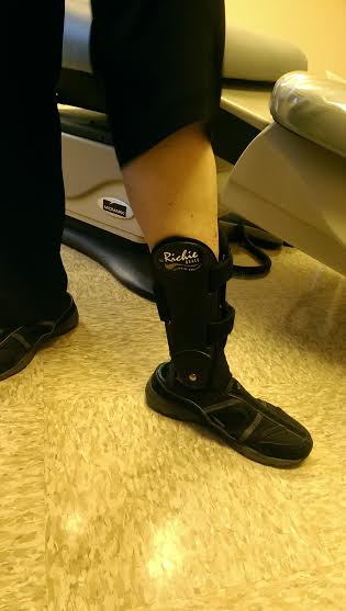
More aggressive stretching can be accomplished using a night splints stretches both the Achilles tendon in the plantar fascia for three or more hours at a time, sometimes all night long.
Ice therapy can reduce the inflammation or two or three hours at a time. I find that the easiest method to apply ice to the heel is to feel a 20 ounce plastic Coke bottle with water and freeze it in the freezer without the cap. Remove the bottle and put the cap back on and roll the midfoot and heel over the depression at the bottom of the Coke bottle. Roll the heel on bottle for 10 min. at a time three times a day. More often is never wrong. You could get in trouble if you apply unclothed skin to ice without motion. The Coke bottle works very well because you roll your heel back and forth on the bottle and never keep the ice in one spot for very long.
Physical therapy:
Physical therapy can often do wonders to control the pain of plantar fasciitis. They will use national level of stretching and taping techniques as well as ultrasound treatments and electrical stimulation to bring the pain under control. They have a technique called electrophoresis and phonophoresis where cortisone preparations are painlessly push through the skin using either gentle electric current or the wand of an ultrasound machine. If you visit either of our physical therapist, Amy or Beth, be prepared to go home with a list of exercises and activities to do on your own to control plantar fascial pain.
Medical:
Pills: The early stages of plantar fasciitis are inflammatory conditions. As such, anti-inflammatory agents can often make life much more bearable. Oral agents like Motrin, Celebrex, Naprosyn and Voltarin can stop the inflammatory process in its tracks. While over-the-counter Motrin can achieve anti-inflammation levels of medication you need to take 2400mg – 3200 mg per day to do that. At 200mg per tablet that is a whopping 12 to 16 tablets a day in divided doses and taken with meals. Few people feel comfortable taking that many tablets so I write for other medications that need to be taken as infrequently as once or twice a day.
Injections: injections of cortisone mixed with a local anesthetic can often be the Holy Grail of heel pain treatment. Many patients have been permanently and forever cured of their heel pain with a single cortisone injection. This fact combined with the near absence of any injection complications makes it the singular best treatment you can have for plantar fasciitis.
Orthotics
If you had a positive response to the low dye strapping you will probably have a positive response to functional orthotics. The orthotics that podiatrist make our oftentimes much better than the ones made by physical therapists or chiropractors. It is a little frustrating to have a patient who has an ineffective pair of orthotics from another practitioner say to us that they “already have orthotics” when we can see from the inserts in their shoes they are nothing like the kinds we make. Even if you have so-called orthotics from other practitioners, if our low dye strapping make sure foot stop hurting so will our functional orthotics in most cases. This is providing, of course, that you’re able to wear the kinds of shoes in which and orthotic is appropriate. Orthotics cannot be worn in most sandals or flip-flops. They also cannot be worn in most high heels but, as I mentioned above, the mere fact of elevating the heel will probably make your heel stop hurting.
To make orthotics in our office we will take either an image or mold of your foot held in what we call the “neutral position” and, and this is important, in a non-weight bearing position. Orthotics made from an outline of your foot or from stepping into foam are not usually considered legitimate orthotics. Once we have your non-weight bearing impression we will send them off to the laboratory along with an order form for one of any number of these signs or styles of orthotic that will hold your foot in this neutral position as you go about your daily activities. Again, I cannot tell you the number of patients who have been, in essence, cured of their plantar fasciitis by wearing a well-made and proper fitting functional orthotics.
Insurance coverage for orthotics
Functional orthotics works so well that it is a shame that Medicare, and some private insurance plans do not cover them. If you want to know if your insurance plan covers functional orthotics we ask that you call them. The phone number should be on the back of your insurance card. Ask them specifically if they cover functional orthotics and give them the code number L3000 which is the medical code number for functional orthotics. Be sure you write down carefully what they say and who you talk to. Also ask if they need any preauthorization prior to paying for functional orthotics. If orthotics are not covered by your insurance, we have a reasonable cash fee.
For more information about orthotics, including the insurance coverage aspect, please call our office and ask for a copy of our “Orthotic Brochure.”
Why do you keep referring to your orthotics as FUNCTIONAL?
From the podiatric point of view orthotics are either functional or accommodative. Accommodative orthotics, often called soft orthotics, are designed just to pad your foot and give it a “cushier” landing. Arch supports, spine levelers and just about anything made of leather or rubber fit into this category. A functional orthotic changes the way your foot functions. It will hold your foot in a pre-designed position. A functional orthotic is made from a plaster or computer impression of your foot, not just measuring the size or having you step into a foam box. Originally accommodative orthotics were made of leather and were designed for patients who needed a more cushioned insole in their shoes. Insurance companies that do cover orthotics require that they be functional to be eligible for coverage.
Who Needs Surgery?
The overwhelming number of cases of plantar fasciitis are brought under control with nonsurgical means.
In our office we offer three types of surgery for resistant plantar fasciitis: 1) shockwave therapy 2)PRP injections and 3) plantar fasciotomy or cutting the plantar fascia.
Shockwave therapy – ESWT.
Sound waves contain energy as anybody who has had their windows rattled by a sonic boom can testify. Shockwave therapy machines gather a very loud shock explosion and concentrated on one tiny little point about 2 inches from the head of the machine. For almost 30 years shockwaves have been used to break apart kidney stones without the need for surgery. For 15 years these little waves have been directed at the insertion site of the plantar fascia on the heel. Unlike what you might expect, attacking a heel spur with sound waves does not break them up. The disappointment at the lack of destruction of the heel spur is offset by the fact this machine has chronic, long-standing plantar fasciitis. The treatment takes about 25 min. and it’s usually done under local anesthesia. After this treatment fully one third of the shockwave patients no longer have any heel pain at all, one third have enough of a reduction that traditional surgery is not needed and one third do not receive much help at all. Surprisingly, there are no known complications from this surgical procedure. Either it helps or it doesn’t but nobody has been made worse. We are one of the only offices in the Southern California area to have a high-energy shockwave therapy machine in our office. We use it for many of the resistant heel cases and it is very effective. The only real complication is financial: many insurances refuse to pay for shockwave therapy. For those of you for whom shockwave therapy is not a covered service we have a very reasonable cash fee structure of $1000 or 1 foot and $1500 if we do both feet. We have another section of our website entirely devoted to shockwave therapy and we ask that you refer to it. https://conejofeet.com/shockwave-therapy
A final note on shockwave therapy, do not confuse the high-energy shockwave therapy that we can offer in our office with the low energy shockwave machines that are found in many other podiatry offices and, especially the entire package and physical therapy offices. High-energy shockwave needs some kind of anesthesia, in most cases. Low energy does not. All of the results we are talking about are based upon high-energy shockwave not low energy. You can suspect that your Dr. is dealing with low energy shockwave if he or she offers you a series of four or six treatments for a fixed fee like $400 or $600. High-energy shockwave therapy is, unfortunately, more expensive than this.
PRP injections
There is a new method of care and is similar to ESWT above except for the plasma is drawn from the blood and injected back into the damaged site. This initiates the healing cascade of healing causing the fascia repair itself.
Plantar Fasciotomy
The most direct and the oldest method for doing surgery on plantar fasciitis is to cut the plantar fascia. The technique involves making a 1-2 inch incision on the medial aspect of the heel and dissecting down to the plantar fascia which is cut. The heel spur does not need to be removed as discussed previously. There are two lesser dramatic plantar fascial surgery procedures: the minimal incision and the endoscopic procedure.
Minimal incisional plantar fasciotomy
The technique involves cutting one third of the plantar fascia to a small, 1/4 inch, incision on the bottom of the heel. This can be performed either in the office or at the surgery Center under local anesthesia with or without a little sedation. The incision takes just one little suture stitches to close and the patient can return to regular shoes in three or four days. This has a remarkable success rate. One of the feared complications of performing a plantar fasciotomy is the possibility of transferring pain to the outside of the foot-the middle of the foot behind the baby toe. Although it is always a risk and always acknowledged on the consent form, it is very unlikely.
Endoscopic plantar fasciotomy
This surgery is done using an endoscope which is a small straw like to which is placed into the heel with a small incision on the inside of the heel. A small, very tiny, camera is placed into the wound and they can actually visualize the plantar fascia as they cut it. This is very elegant. Because this is done at the surgery center under local anesthesia it cannot be done in the office.
Who should do the surgery?
There are only two categories of doctors who commonly and routinely perform plantar fasciotomy and heel spur surgeries: podiatrist and orthopedic surgeons. In your area you should use the professional who does the most plantar fascial surgeries and has the most experience. While that could be an orthopedic surgeon in most areas very few orthopedic surgeries do very many foot surgeries. If you are bidding surgeon says he or she does a lot of heel surgeries then you are probably in good hands. More than likely, however, you will be getting surgery from a local podiatrist. And if you do choose the podiatrist, we recommend that you pick one that is Board Certified by the American Board of Foot and Ankle Surgery. You can usually identify these surgeons because they are also Fellows of the American College of Foot and Ankle Surgeons which means they have the initials FACFAS after their name. In our office all three doctors are Fellows of the American College of Foot and Ankle Surgeons
Thank you for reading this far. If you have questions that I could answer, feel free to leave them on https://conejofeet.com


3 thoughts on “A Patient’s Guide to Heel Pain”
Great article! Thanks for the good info. Seeing Dr Payne next week
Glad you found the article helpful Mike, make sure to check out all the other articles we have on the website. Dr. Zapf is very glad to have another avenue of communication with our patients. Keeping reading, and see you soon!
-Dr. Darren Payne